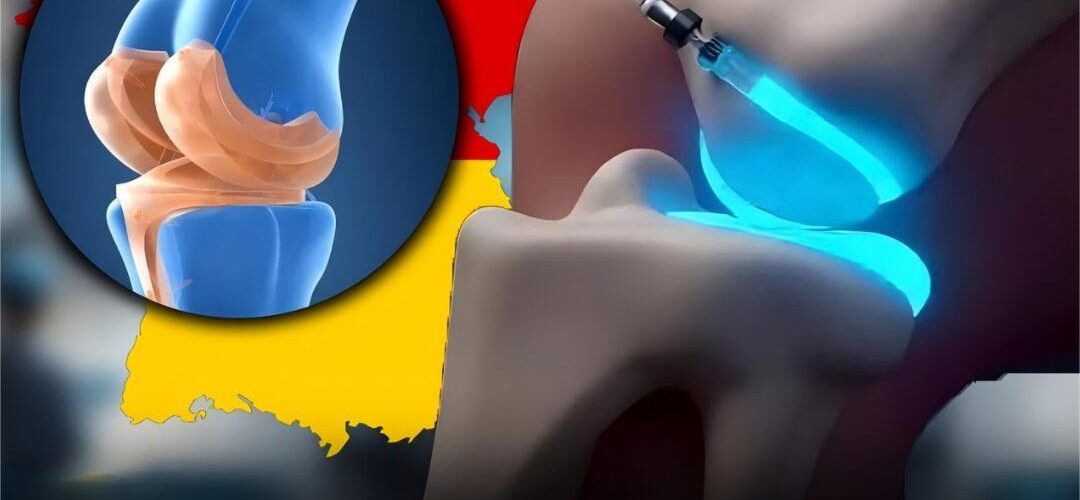
Berlin, Deutschland – Kürzlich verbreitete sich in den sozialen Medien die Nachricht, deutsche Wissenschaftler hätten ein „Wundergel“ erfunden, das abgenutzten Knorpel durch eine einfache Injektion nachwachsen lassen und so invasive Operationen überflüssig machen könne. Obwohl die Realität komplexer ist, spiegelt die Geschichte echte Fortschritte in der Biomaterial- und Regenerativen Medizin wider , die eines Tages die Behandlung von Arthritis und Gelenkverschleiß revolutionieren könnten.
Das Versprechen der Knorpelregeneration
Knorpel, das glatte Gewebe, das die Gelenke polstert, galt lange Zeit als nahezu unheilbar, wenn er einmal beschädigt ist. Im Gegensatz zu Haut oder Knochen hat er keine Blutgefäße und nur eine geringe natürliche Heilungsfähigkeit. Für Millionen von Arthritispatienten bedeutet Knorpelverlust chronische Schmerzen, eingeschränkte Beweglichkeit und oft die Notwendigkeit einer Gelenkersatzoperation.
Wissenschaftler auf der ganzen Welt suchen nach Möglichkeiten zum Wiederaufbau von Knorpel und biotechnologisch hergestellte Gele und Gerüste haben sich als einer der vielversprechendsten Ansätze herausgestellt.
Der deutsche Beitrag: ChondroFiller®
In Deutschland entwickelten Forscher ChondroFiller® , ein 2013 eingeführtes Gel auf Kollagenbasis. Das Gel wird mittels minimalinvasiver Arthroskopie und nicht durch eine einfache Injektion mit einer Spritze verabreicht. Es füllt Knorpeldefekte und dient als Gerüst für neues Gewebewachstum.
Sobald das Gel eingebracht ist, hilft es den körpereigenen Zellen, den beschädigten Bereich zu reparieren. Die Patienten müssen sich einer standardmäßigen postoperativen Behandlung unterziehen, die eine kurze Ruhigstellungsphase und Physiotherapie umfasst. Wichtig ist, dass ChondroFiller® in Europa seit über einem Jahrzehnt zugelassen ist, aber es handelt sich nicht um den „neuen Durchbruch“, der manchmal in viralen Posts suggeriert wird.
Neue Hydrogelforschung
Neben ChondroFiller® experimentieren Wissenschaftler auch mit Hydrogelen der nächsten Generation , die die Knorpelregeneration effektiver stimulieren sollen. Eine aktuelle Studie in Nature Communications (2023) beschrieb ein „intelligentes“ Hydrogel, das therapeutische Moleküle schrittweise freisetzen und gleichzeitig seine Form an Gelenkdefekte anpassen kann. In Tierversuchen förderte dieses Gel das Knorpelwachstum , verbesserte die Gelenkfunktion und reduzierte Narbenbildung.
Obwohl sich derartige Innovationen noch in der präklinischen Phase befinden, deuten sie auf eine Zukunft hin, in der minimalinvasive Geltherapien Gelenkersatzoperationen ersetzen oder verzögern könnten.
Zwischen Hype und Realität
Während in den sozialen Medien die Behauptungen über eine neue deutsche „Heilung nur durch Injektion“ verstärkt werden, weisen Experten darauf hin, dass es derzeit keine solche Behandlung auf dem Markt gibt. Im Moment:
- ChondroFiller® ist real, erfordert aber eine arthroskopische Platzierung.
- Die Hydrogelforschung schreitet voran, befindet sich jedoch noch im Stadium von Labor- oder Tierstudien.
- Für eine einfache Injektion, die den Knorpel vollständig wiederherstellt, gibt es noch keinen klinischen Beweis .
Dr. Anne Müller, Spezialistin für Regenerative Medizin in Berlin, stellt fest:
„Die Wissenschaft entwickelt sich rasant, aber Patienten sollten verstehen, dass die Knorpelregeneration eine der größten Herausforderungen in der Orthopädie darstellt. Wir sind näher dran als je zuvor, aber wir haben die Ziellinie noch nicht erreicht.“
Ein Blick in die Zukunft
Wenn die laufenden Versuche erfolgreich sind, könnten bioaktive Gele letztendlich Folgendes bieten:
- Nicht-invasive Alternativen zu größeren Operationen.
- Schnellere Genesungszeiten und weniger Narbenbildung.
- Verbesserte Mobilität und Schmerzlinderung für Millionen von Osteoarthritis-Patienten.
Bis dahin müssen sich die Patienten auf etablierte Behandlungsmethoden verlassen, während die Forscher weiterhin versuchen, die Fähigkeit des Körpers zur Heilung seiner Gelenke zu entschlüsseln.
Fazit:
Die deutsche Arbeit an Knorpelreparaturgelen verdeutlicht sowohl die Verheißung als auch die Herausforderungen der regenerativen Medizin. Während aktuelle Produkte wie ChondroFiller® echte Vorteile bieten und experimentelle Hydrogele spannende Möglichkeiten eröffnen, ist die virale Behauptung einer einfachen Injektion, die „Knorpel ohne Operation nachwachsen lässt“, eher Hype als Realität .
Dennoch ist der Tag, an dem sich die orthopädische Versorgung vom Ersatz zur Regeneration verlagert, möglicherweise nicht mehr fern.
37,368 HFMD cases logged nationwide
MANILA, Philippines — The Department of Health (DOH) has reported a sharp rise in hand, foot and mouth disease (HFMD) cases nationwide, with figures showing a nearly 750 percent increase compared to last year.
📈 HFMD Cases Spike Among Young Children
DOH data showed that as of August 9, there have been 37,368 HFMD cases across the country since January — far higher than the 5,081 cases recorded during the same period in 2024.
Health officials noted that most of the patients are children aged one to three years old, consistent with the disease’s prevalence among children under five.
While HFMD is generally mild and self-limiting, the DOH warned that complications such as meningitis, encephalitis, and polio-like paralysis can occur if left untreated.
🦠 Transmission and Symptoms
HFMD spreads easily through direct contact with infected individuals, as well as through droplets and contaminated objects.
Common symptoms include:
- Fever lasting one to two days
- Painful mouth sores
- Rashes and blisters on the hands, feet, and buttocks
Patients are advised to undergo isolation for seven to 10 days until fever subsides and rashes disappear.
The DOH emphasized the importance of strict hygiene practices:
- Utensils and personal items used by the patient must be kept separate and disinfected.
- The isolation room must be thoroughly cleaned once the patient recovers.
Health Secretary Ted Herbosa underscored the need to teach children proper handwashing techniques, especially if they have been exposed to HFMD cases.
💡 Preventive Measures
To reduce the spread of HFMD, health experts recommend:
- Frequent handwashing with soap and water
- Regular disinfection of frequently touched surfaces
- Wearing protective gear (masks, gloves, gowns) when caring for an infected patient
🌧️ Leptospirosis Cases Decline
In contrast, leptospirosis cases — which surged following recent tropical storms — are now on a downward trend.
Only 18 cases were reported nationwide from August 17 to 21, a significant drop from the 1,112 cases recorded in the week following storms Crising, Dante, and Emong earlier this month.
Since the rainy season officially began on June 8, the country has reported 4,436 leptospirosis cases.
State-run hospitals have noted declining admissions:
- Tondo Medical Center reported seven new cases.
- National Kidney and Transplant Institute recorded just one.
- East Avenue Medical Center reported none.
To ensure swift care, the DOH has established fast lanes in hospitals specifically for leptospirosis patients.
✅ Outlook
The DOH continues to monitor both HFMD and leptospirosis closely, urging the public to remain vigilant. While HFMD cases climb at an alarming rate, the recent decline in leptospirosis offers a reminder that strict prevention and early intervention are key to controlling infectious disease outbreaks.
High cholesterol can be inherited. Here’s what you can do about it
Most people associate high cholesterol with an unhealthy diet, lack of exercise, or aging — something that builds up slowly over time. But for some, elevated cholesterol isn’t the result of lifestyle at all. Instead, it’s inherited through a genetic condition known as familial hypercholesterolemia (FH).
🧬 What Is Familial Hypercholesterolemia?
FH is caused by a genetic mutation that leads to lifelong high LDL (“bad”) cholesterol levels, regardless of diet or activity. According to the U.S. Centers for Disease Control and Prevention (CDC), about 1 in 250 people has FH. Yet experts estimate that 70–90 percent remain undiagnosed, leaving them at risk for serious heart problems.
Without proper management, FH greatly increases the risk of early heart attacks and strokes. Detecting it early can be lifesaving.
🩺 Why Cholesterol Matters
Cholesterol itself isn’t harmful — in fact, it’s essential. Every cell in the body needs cholesterol to build membranes and produce hormones. But problems arise when LDL cholesterol builds up in the artery walls.
Excess LDL particles trigger inflammation, much like a splinter under the skin. Over time, this can damage artery linings and cause dangerous blood clots — the underlying process behind most heart attacks and strokes.
⚠️ Signs and Symptoms
Most people with high cholesterol feel no symptoms until serious complications occur. FH is especially insidious because cholesterol levels are high from birth.
In rare cases, very high cholesterol may cause:
- Fatty deposits in tendons (xanthomas)
- Yellowish patches on the skin
- A white ring around the cornea of the eye
But for most, routine blood tests are the only way to detect it.
🔎 Who Should Be Tested?
- Children: Pediatric guidelines recommend cholesterol screening between ages 9–11 if a parent has FH. Some children may be tested earlier.
- Adults: There’s no age “too early” for testing. Anyone with a family history of heart disease, stroke, or high cholesterol should ask for screening.
It’s also important to test for lipoprotein(a), another inherited cholesterol particle often elevated in people with FH, which further increases cardiovascular risk.
🥗 Living With Inherited High Cholesterol
Lifestyle changes alone won’t normalize cholesterol in FH — but they still matter. According to cardiologists, healthy habits can cut cardiac risk by up to 80 percent, even when LDL remains high.
Recommendations include:
- Eating fruits, vegetables, whole grains, lean protein, and nuts
- Limiting added sugars, sodium, red meat, and processed foods
- Exercising at least 150 minutes of moderate activity per week (walking, cycling, swimming)
- Avoiding smoking entirely
Most people with FH will also need cholesterol-lowering medications, such as statins or newer therapies, to reduce LDL and protect the heart.
✅ A Winnable Battle
A diagnosis of familial hypercholesterolemia may sound daunting, but experts emphasize that it’s treatable and manageable. With the right combination of medical care, lifestyle adjustments, and early detection, people with FH can live long, healthy lives.
As cardiologist Dr. Ashish Sarraju explains:
“In most cases, I consider high cholesterol treatable and a winnable battle.”
The key is awareness — recognizing that if heart disease runs in your family, testing early could make all the difference.
Groundbreaking Surgery Removes Spinal Tumor Through the Eye Socket
In a remarkable medical first, surgeons at the University of Maryland Medical Center have successfully removed a spinal tumor by accessing it through a patient’s eye socket. The pioneering procedure marks a new milestone in neurosurgery, offering fresh hope to patients facing rare and life-threatening tumors.
A Rare and Dangerous Diagnosis
The patient, 19-year-old Karla Flores, was diagnosed with a chordoma—a rare tumor that arises in the bones of the skull and spine. Although slow-growing, chordomas are highly invasive and often wrap around delicate structures such as nerves, arteries, and the spinal cord.
Flores’ tumor was lodged in her cervical spine, perilously close to critical blood vessels and nerves. Conventional surgery posed extraordinary risks, including paralysis, loss of vital functions, or even death. The location made traditional approaches nearly impossible without causing severe damage.
A Revolutionary Surgical Pathway
Faced with these challenges, neurosurgeon Dr. Mohamed A.M. Labib and his team adopted a bold approach. Rather than using large incisions, they employed a transorbital route—entering through the natural opening of the eye socket.
This technique, previously used in select brain tumor operations, was adapted for spinal surgery after extensive research. Surgeons created a precise corridor through the orbit, guided by advanced imaging, navigation systems, and specialized instruments.
The outcome was extraordinary: the tumor was removed, essential neurological functions were preserved, and the patient was left with no external scarring.
A Team Effort
The achievement was the result of collaboration across multiple specialties, including neurosurgery, head and neck surgery, radiation oncology, and spinal reconstruction. After the tumor removal, Flores received proton radiation therapy—a highly targeted treatment designed to minimize collateral damage—as well as spinal fusion to stabilize her neck.
Recovery and Inspiration
Today, Flores is cancer-free and regaining strength. Her case has been hailed as a breakthrough in minimally invasive neurosurgery.
“This is the first step in a new direction,” said Dr. Labib. “Areas of the spine once considered too dangerous to operate on may now be accessible with less invasive methods.”
Redefining the Future of Neurosurgery
The success of this transorbital approach could transform how surgeons treat complex spinal and skull base tumors. By reducing trauma, shortening recovery times, and avoiding disfiguring scars, it may become an invaluable option for conditions once thought untreatable.
Experts believe the method could one day be expanded to treat other spinal and cranial conditions, pushing the boundaries of what modern medicine can achieve.
A New Era in Surgical Innovation
For Flores, the operation was life-saving. For the medical community, it represents a new era—where procedures once deemed impossible can now be performed with precision and minimal invasiveness.
As more hospitals explore and refine this approach, the promise is clear: safer surgeries, faster recoveries, and renewed hope for patients facing the most complex of diagnoses.